Light Sheet Microscopy Research
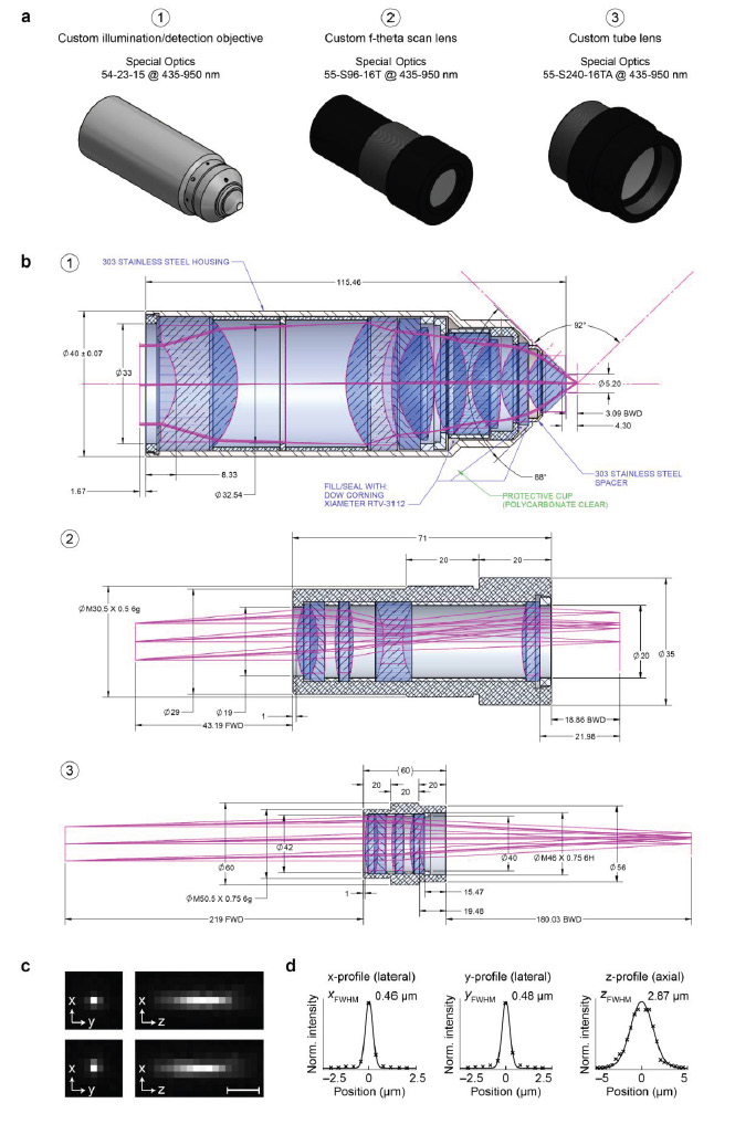
Special Optics designed and developed a set of three custom lenses used in the new light-sheet microscope developed by Philipp Keller and Raghav Chhetri of Howard Hughes Medical Institute's Keller Lab in 2015. The IsoView microscope produces three-dimensional images of entire organisms within less than a second. The IsoView incorporates highly specialized microscope objectives, f-theta scan lenses and illumination tube lenses from Special Optics. This unique approach and innovative design overcomes one of the most common limitations in light microscopy - poor spatial resolution along the axial dimension when viewing samples from multiple angles.
Images of a sample are collected using four custom illumination/detection objectives positioned at right angles to one another – similar to traffic lights at an intersection. Each objective sends light into the sample to illuminate it, and also collects fluorescent light emitted by the sample.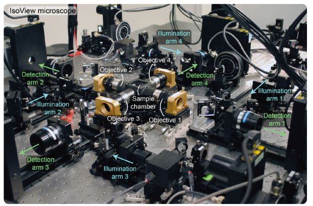
Core of IsoView light-sheet microscope developed by Phillip Keller and Raghav Chhetri. Image courtesy of and used with permission from Raghav Chhetri.
From each side, an objective produces a thin beam of light that sweeps the sample from top to bottom so quickly that the detection camera across from it sees a continuous sheet of light. The beams from each of the four objectives are staggered so that they do not interfere or intersect with one another, and a rolling shutter in each camera keeps pace with the beam, so that the camera's detector remains focused on the narrow slit along which it can pick up high-resolution information. These features allow the microscope to collect four different images of the sample from different angles simultaneously, without any crosstalk between these multiple views.
 The objectives allow for precise control over the focus of the illumination and collection of light and image that is relayed to the cameras. In the microscope Keller and Chhetri utilized the grouping of the objective, scan and tube lens and created four separate channels – allowing for four-view and two-color imaging
-
Custom Illumination/Detection Objective:
Special Optics part number: 54-23-15. Transmittance/Wavelength: 435-950nm, f= 15 mm, NA = 0.714 (water) -
Custom Telecentric F-theta Scan Lens (used within the Illumination sub-system):
Special Optics part number: 55-S96-16T. Transmittance/Wavelength: 435-950nm -
Custom Tube Lens (used within detection sub-system):
Special Optics part number: 55-S240-16TA. Transmittance/Wavelength: 435-950nm